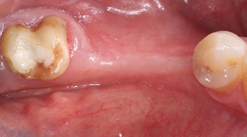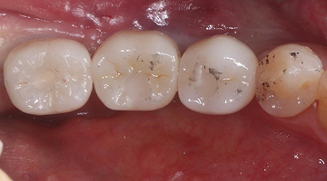
ضرسان بعد سبع سنوات: علاج عصب بدون تلبيس، وحشو يحافظ على العصب
ضاحك علوي احتاج علاج عصب فأعدنا بناءه وغطّيناه بحشو تجميلي بدل التلبيسة، وضرس سفلي بتسوس عميق أزلناه جزئيًا لنحافظ على العصب حيًّا. والاثنان سليمان بعد سبع سنوات.
من الصف الخامس الابتدائي… إلى زميلة في المهنة
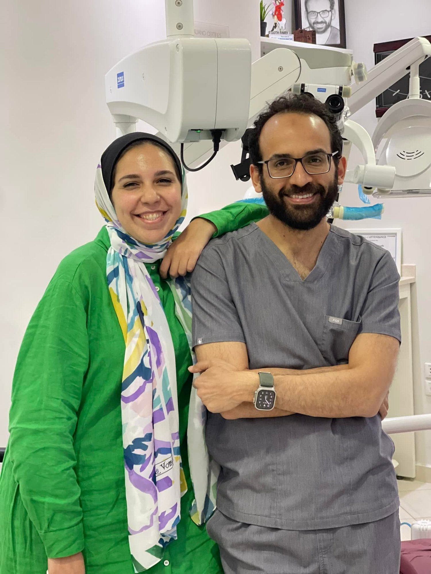
أختي د. نورهان بدأت علاج أسنانها معي وهي في الصف الخامس الابتدائي، وهي اليوم طبيبة أسنان. على مدار السنين عالجت لها كل شيء تقريبًا: علاج جذور، وعلاج لثة، وزراعة، وحشوات تجميلية، وتيجان.
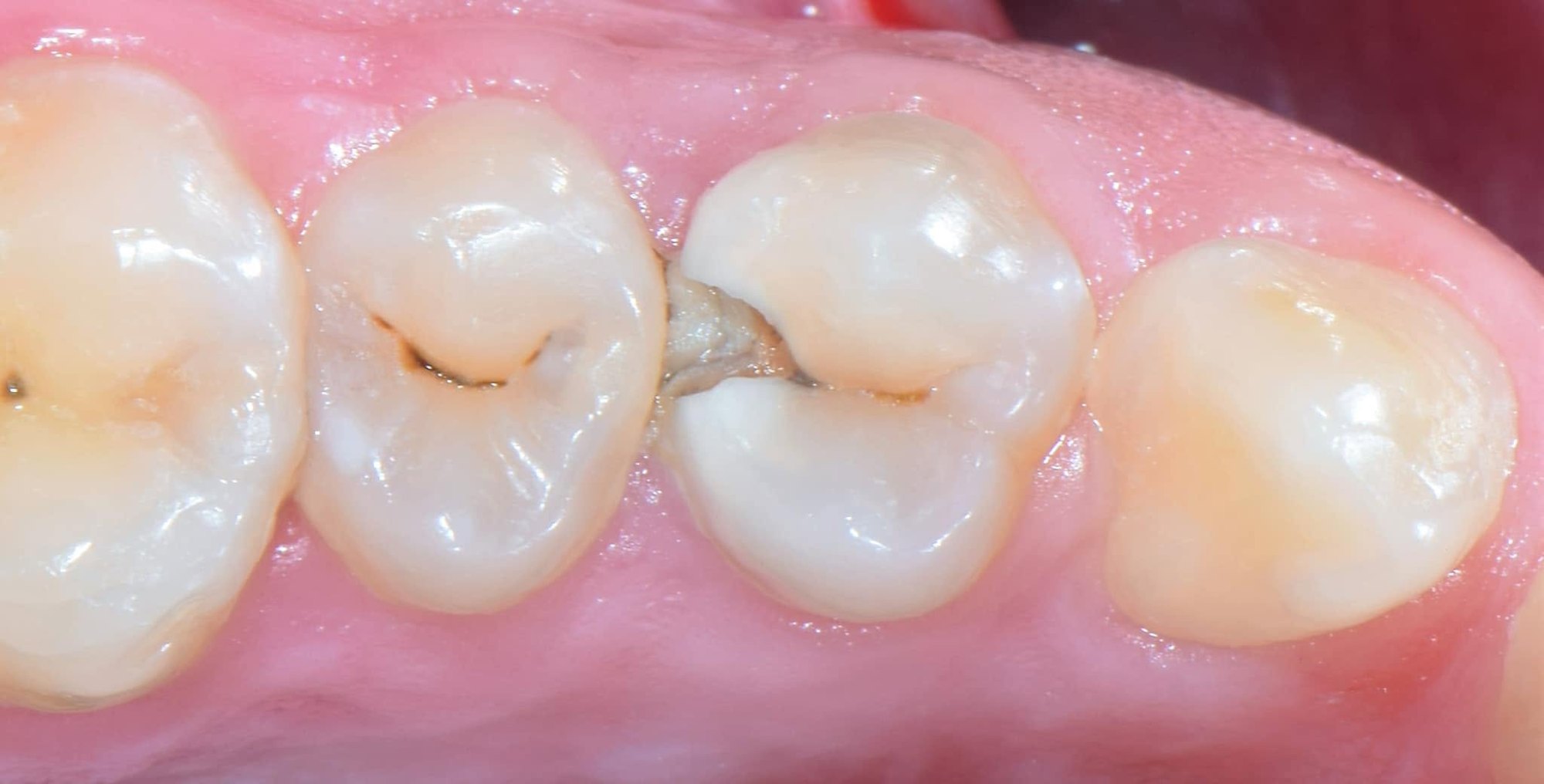
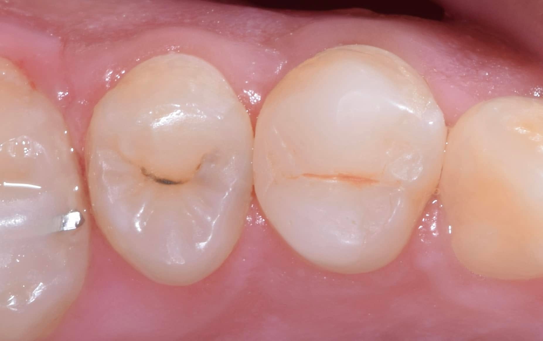
قبل سبع سنوات احتاج ضرسان عندها إلى علاج في الوقت نفسه، ولكل منهما قصة مختلفة.
الضاحك العلوي: علاج عصب بدون تلبيسة
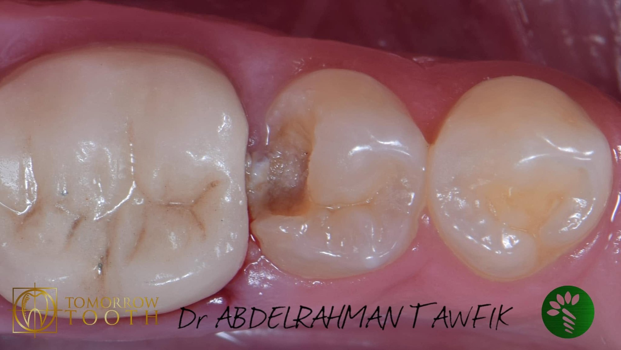
كان التسوس في الضاحك الأول العلوي الأيسر عميقًا حتى أسفل حدود اللثة، ووصل إلى العصب. والمعتاد في حالة كهذه أن يُبرد السن ويُركَّب له تاج (تلبيسة).
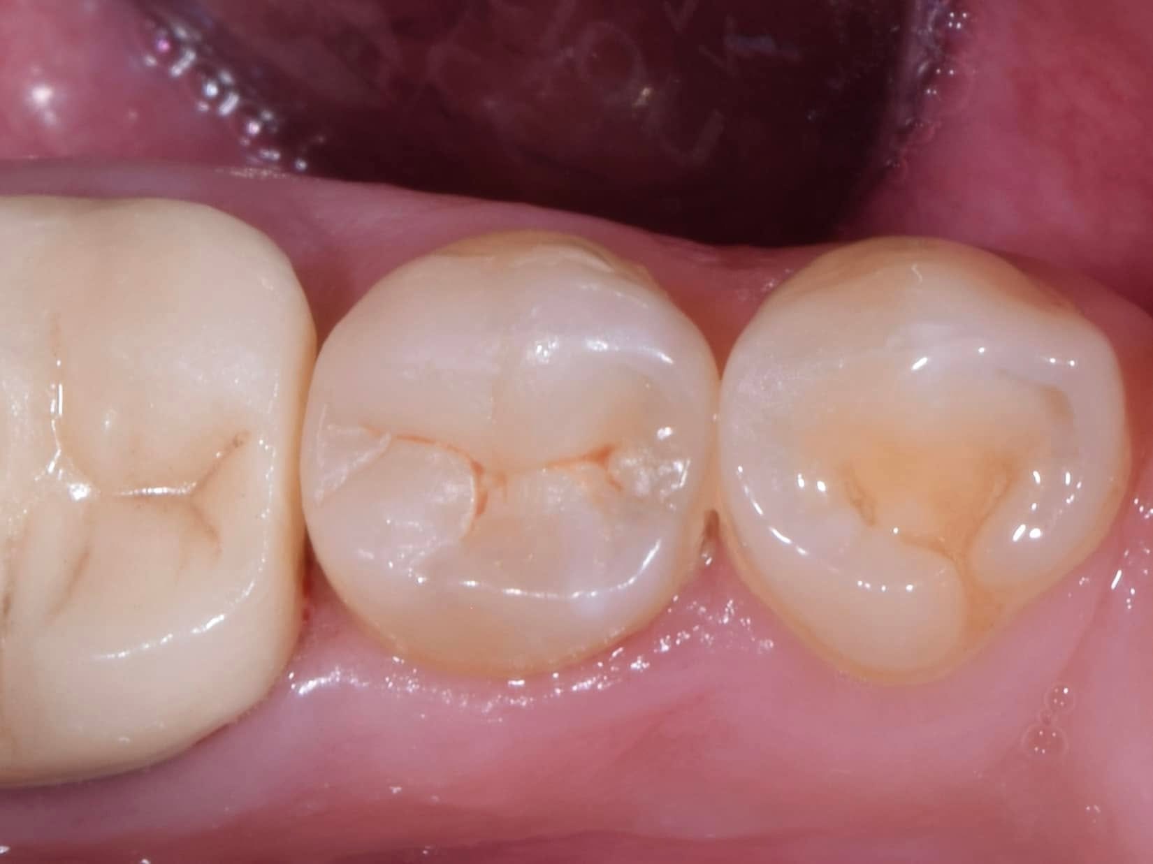
اخترنا طريقًا يحافظ على السن الطبيعي: أعدنا بناء الجزء المفقود من السن قبل علاج العصب، ثم أكملنا علاج العصب، وبعده غطّينا السن بحشو تجميلي مباشر يحمي أطرافه من الكسر، دون برد أي جزء سليم منه.
الضرس السفلي: حشو يحافظ على العصب حيًّا
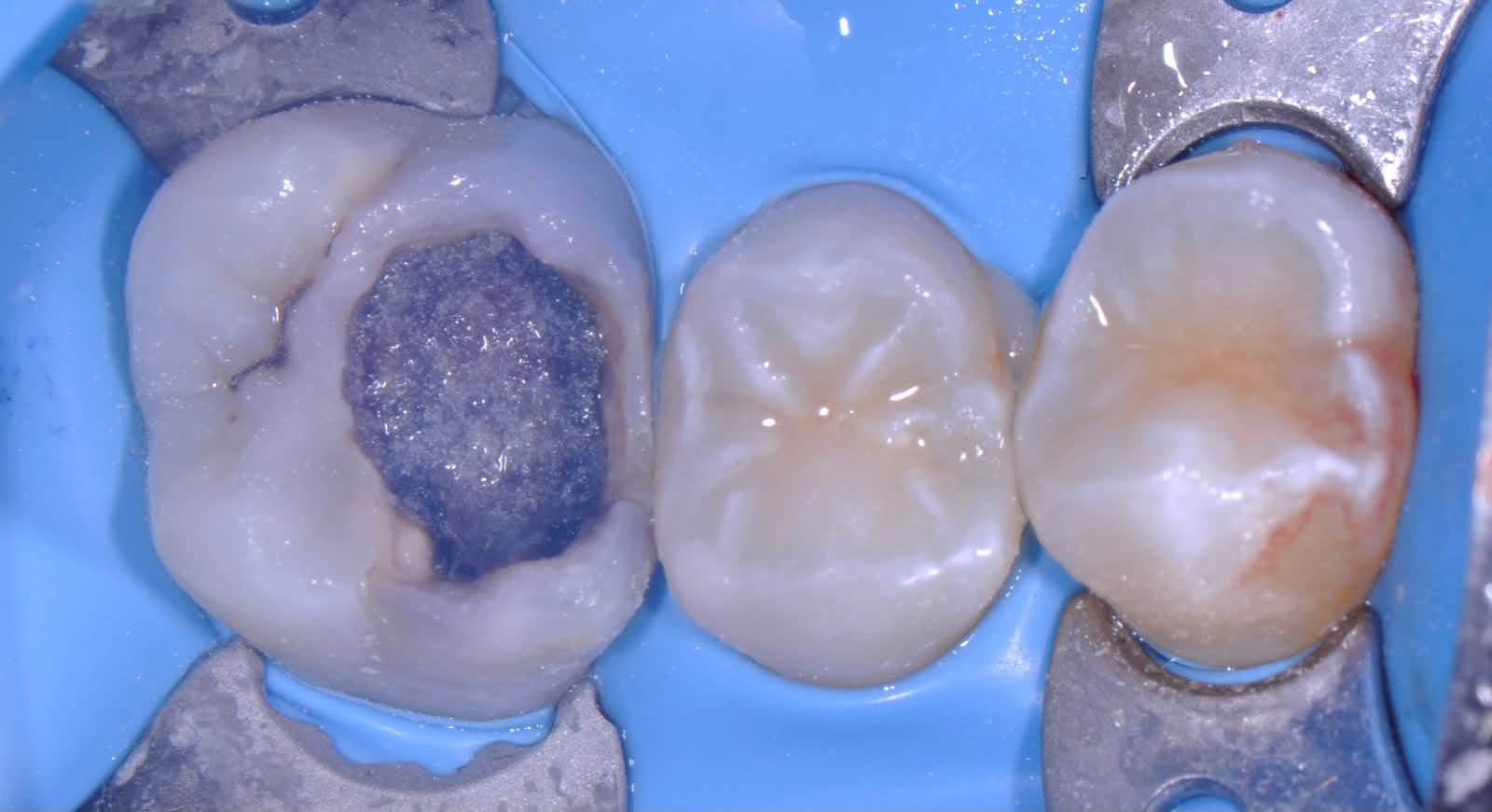
الضرس السفلي كان فيه تسوس عميق قريب جدًا من العصب. لو أزلنا كل التسوس حتى آخره، كان العصب سينكشف ويحتاج الضرس إلى علاج عصب.
لذلك أزلنا التسوس إزالة جزئية مدروسة: نظّفنا الأطراف تمامًا، وتركنا طبقة رقيقة فوق العصب، ثم أغلقنا الضرس بحشو محكم. بهذا بقي العصب حيًّا، وتجنّبنا علاج العصب من الأساس.
بعد سبع سنوات
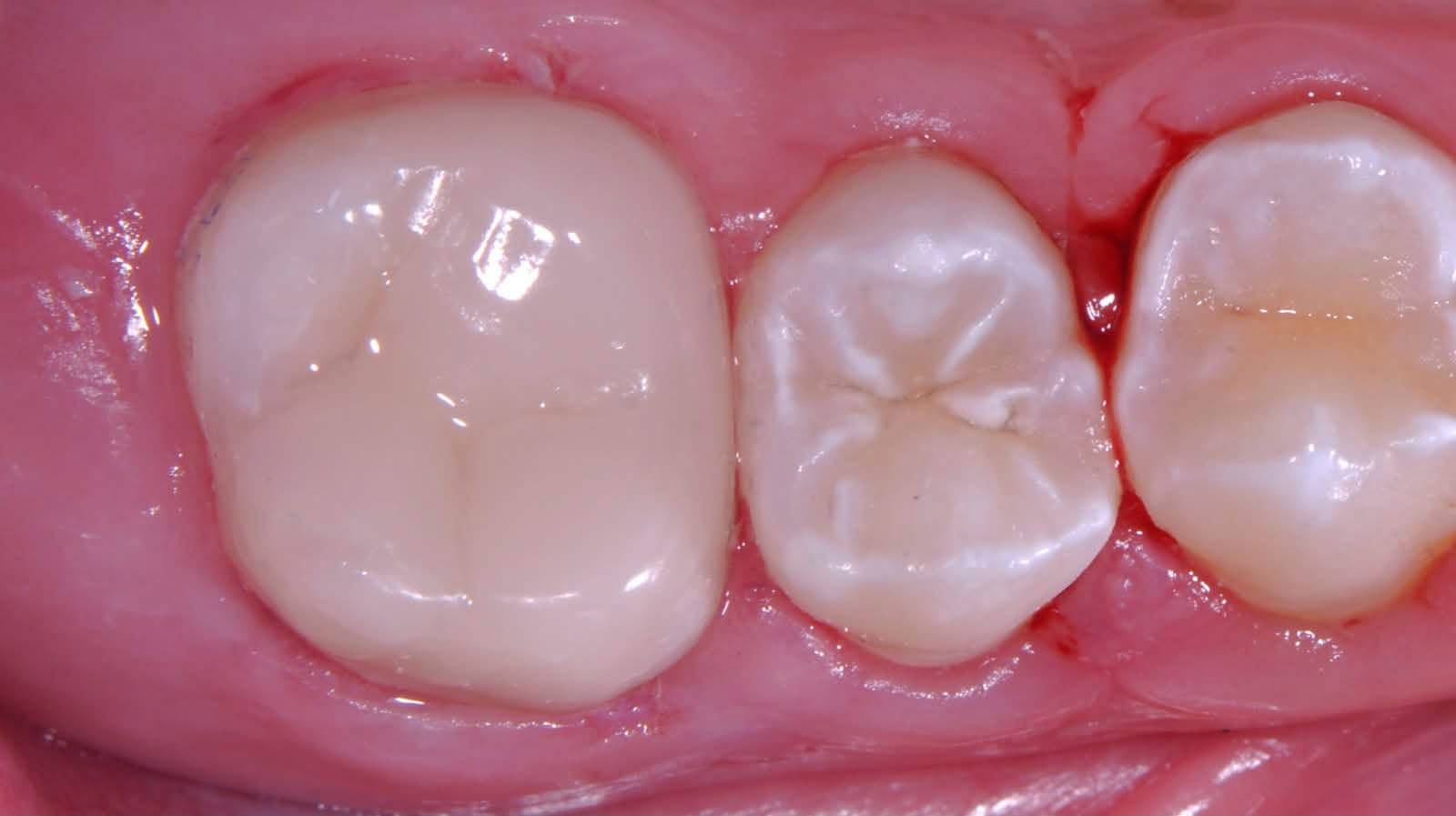
اليوم، بعد سبع سنوات، الحشوان في مكانهما، والضرسان سليمان، واللثة حولهما صحية. والضرس السفلي ما زال حيًّا بعصبه الطبيعي.
أسئلة شائعة
- هل يجب تركيب تلبيسة بعد علاج العصب دائمًا؟ ليس دائمًا؛ إذا بقي جزء كافٍ من السن يمكن حمايته بحشو يغطي أطرافه.
- هل يمكن تجنّب علاج العصب إذا كان التسوس عميقًا؟ في حالات كثيرة نعم، إذا كان العصب سليمًا ولا يوجد ألم مستمر. الإزالة الجزئية للتسوس تحافظ على العصب حيًّا.
- كم يعيش الحشو التجميلي في الأسنان الخلفية؟ أغلب الحشوات تستمر أكثر من عشر سنوات مع العناية والمتابعة الدورية.
- هل الإجراء مؤلم؟ يتم تحت تخدير موضعي، ولا يشعر المريض بألم أثناء العلاج.
التفاصيل الإكلينيكية للأطباء
1 Upper premolar: pre-op, deep proximal caries below the gum line 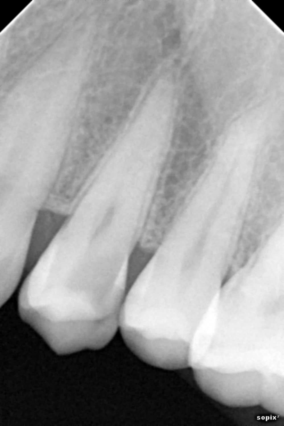
2 Upper premolar: pre-operative radiograph 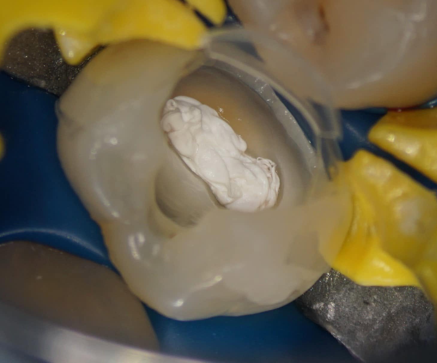
3 Upper premolar: rubber dam and pre-endodontic build-up 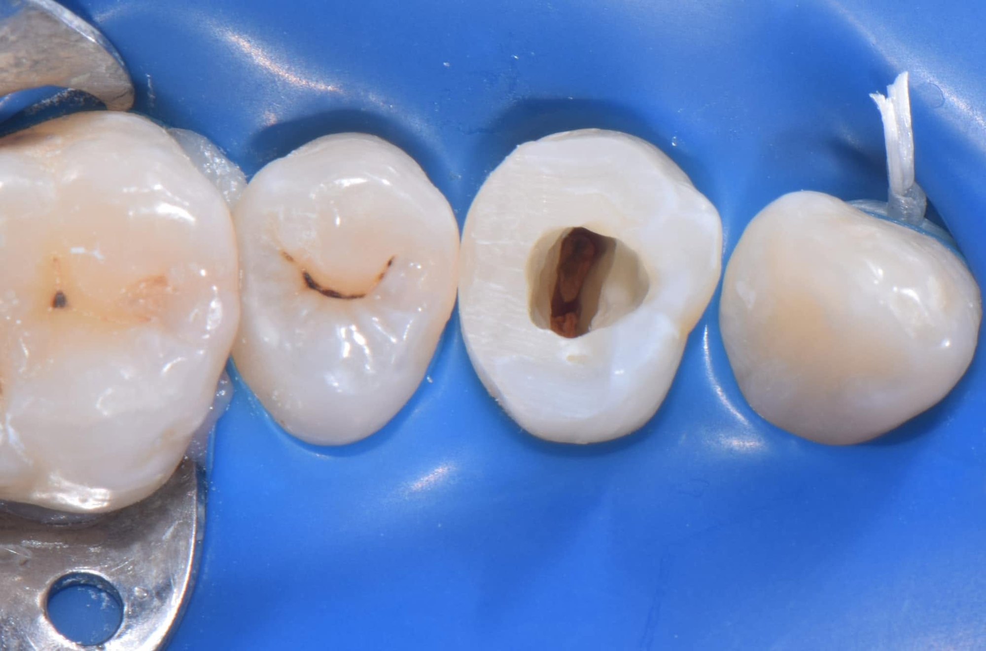
4 Upper premolar: access cavity after the build-up 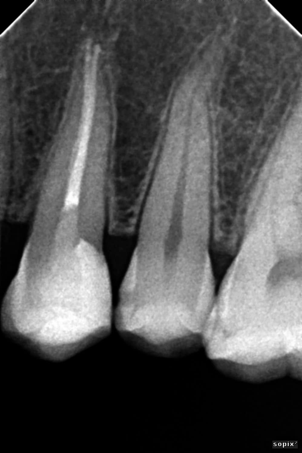
5 Upper premolar: root canal obturation (radiograph) 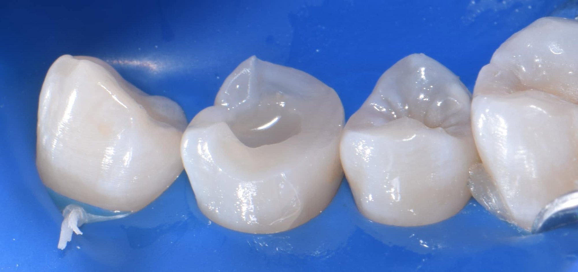
6 Upper premolar: direct cuspal-coverage composite under rubber dam 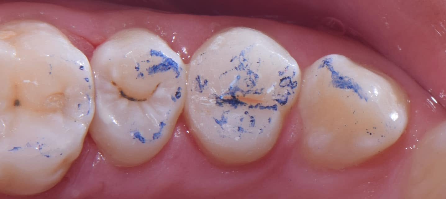
7 Upper premolar: occlusion check 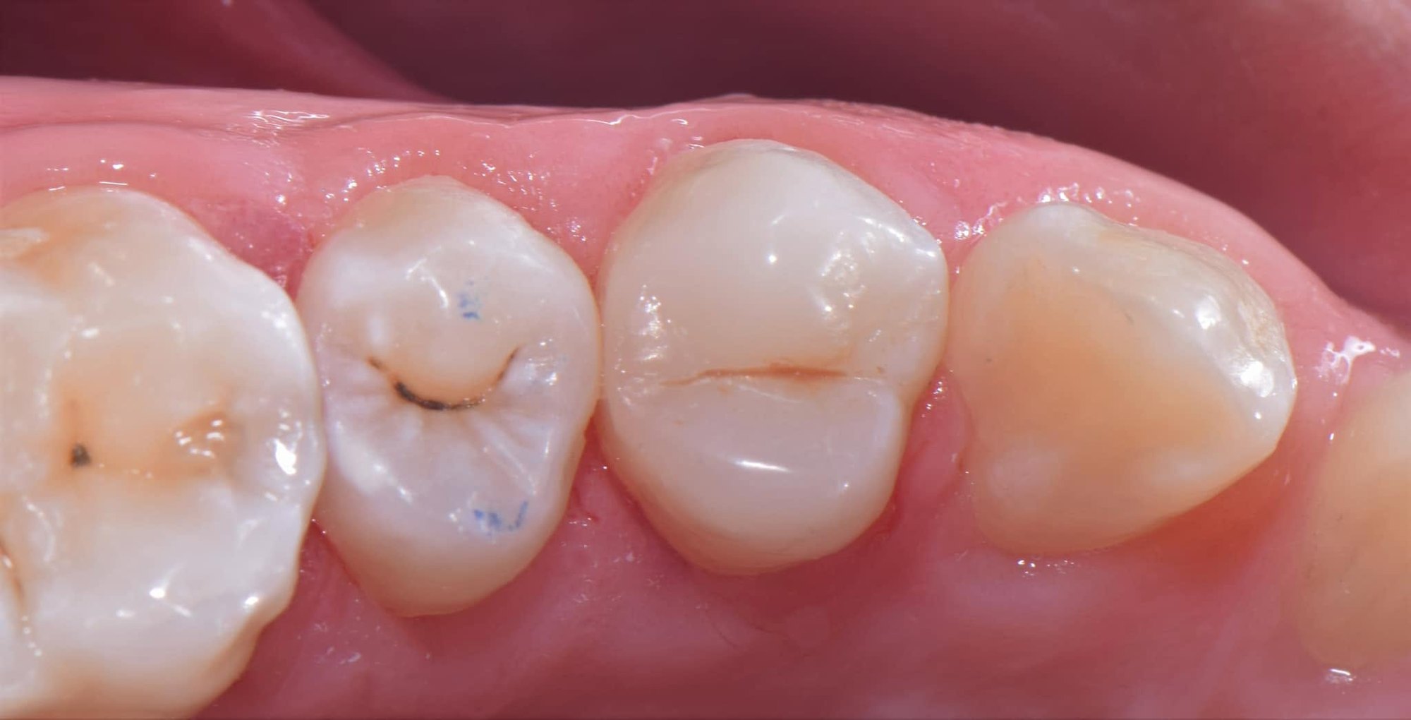
8 Upper premolar: final restoration 9 Upper premolar: 7-year follow-up 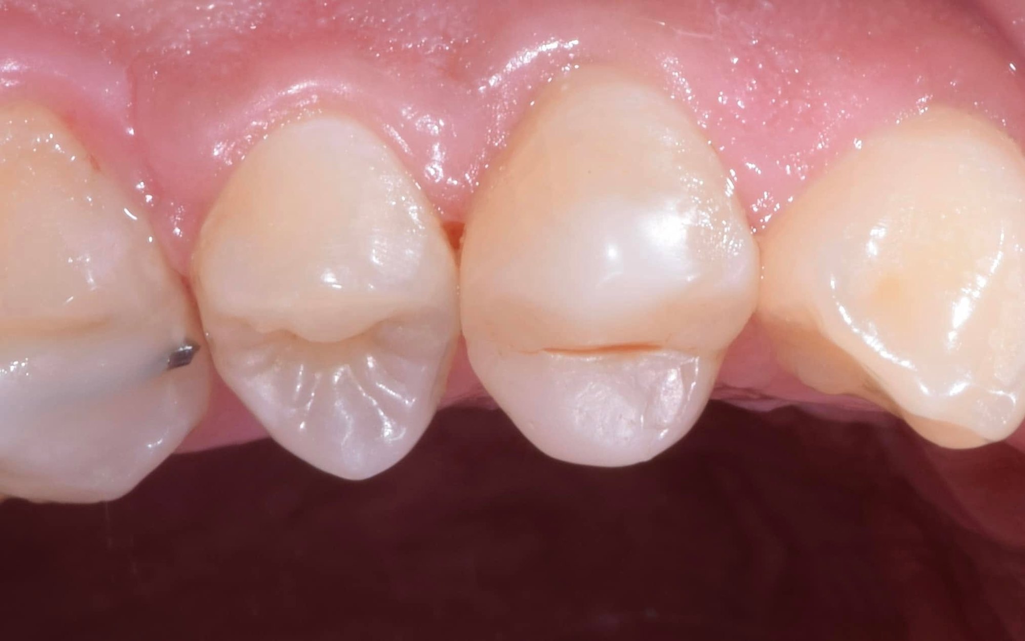
10 Upper premolar: 7-year follow-up 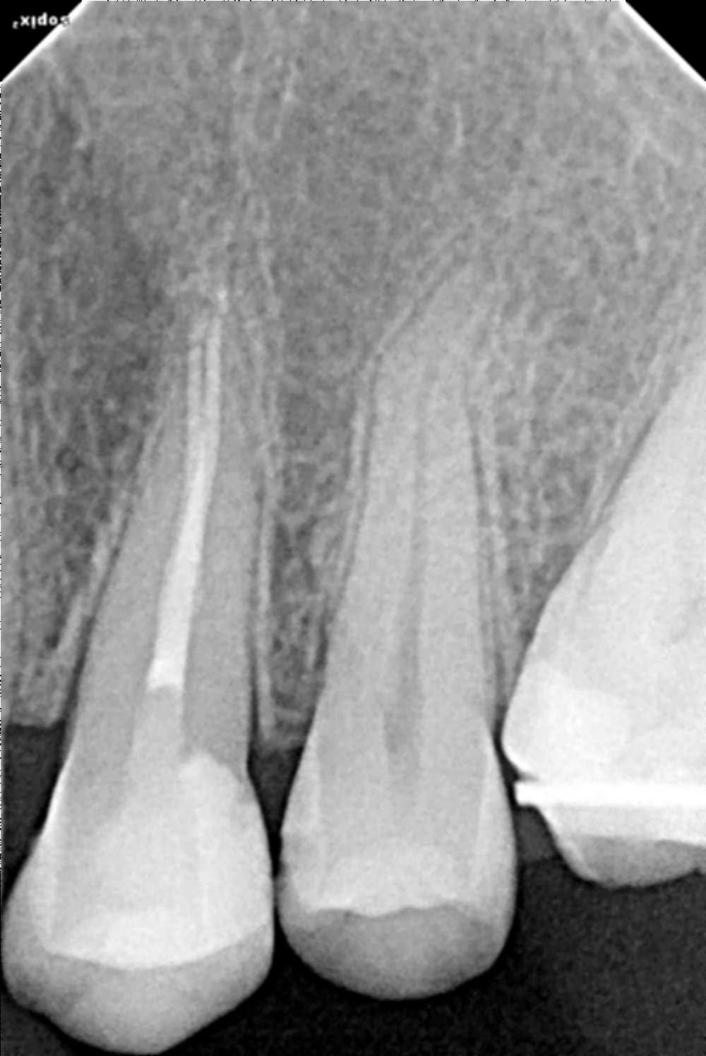
11 Upper premolar: 7-year follow-up radiograph 12 Lower molar: pre-op, deep caries close to the pulp 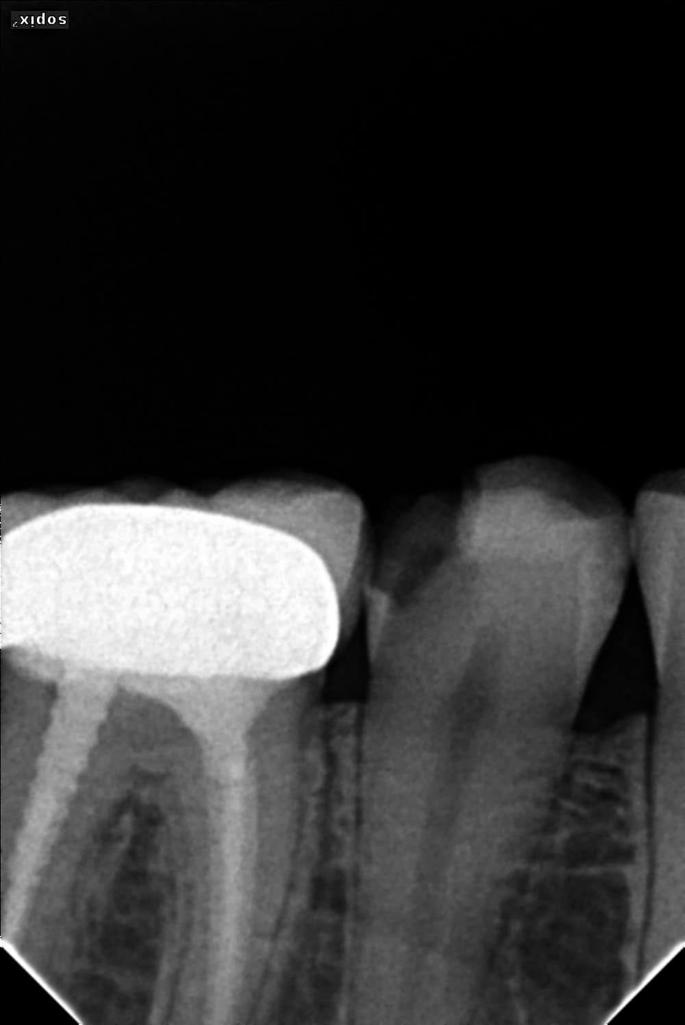
13 Lower molar: pre-operative radiograph 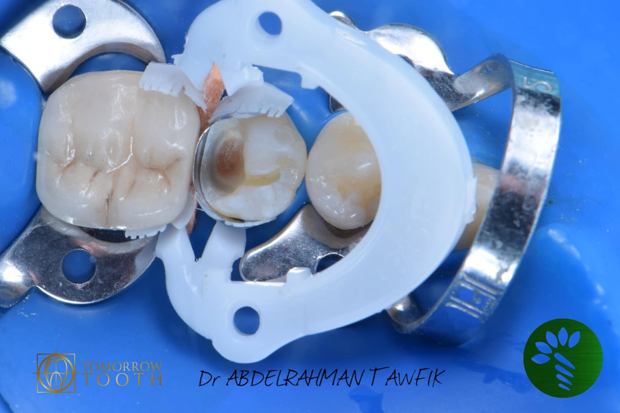
14 Lower molar: multiple-tooth isolation, partial caries removal 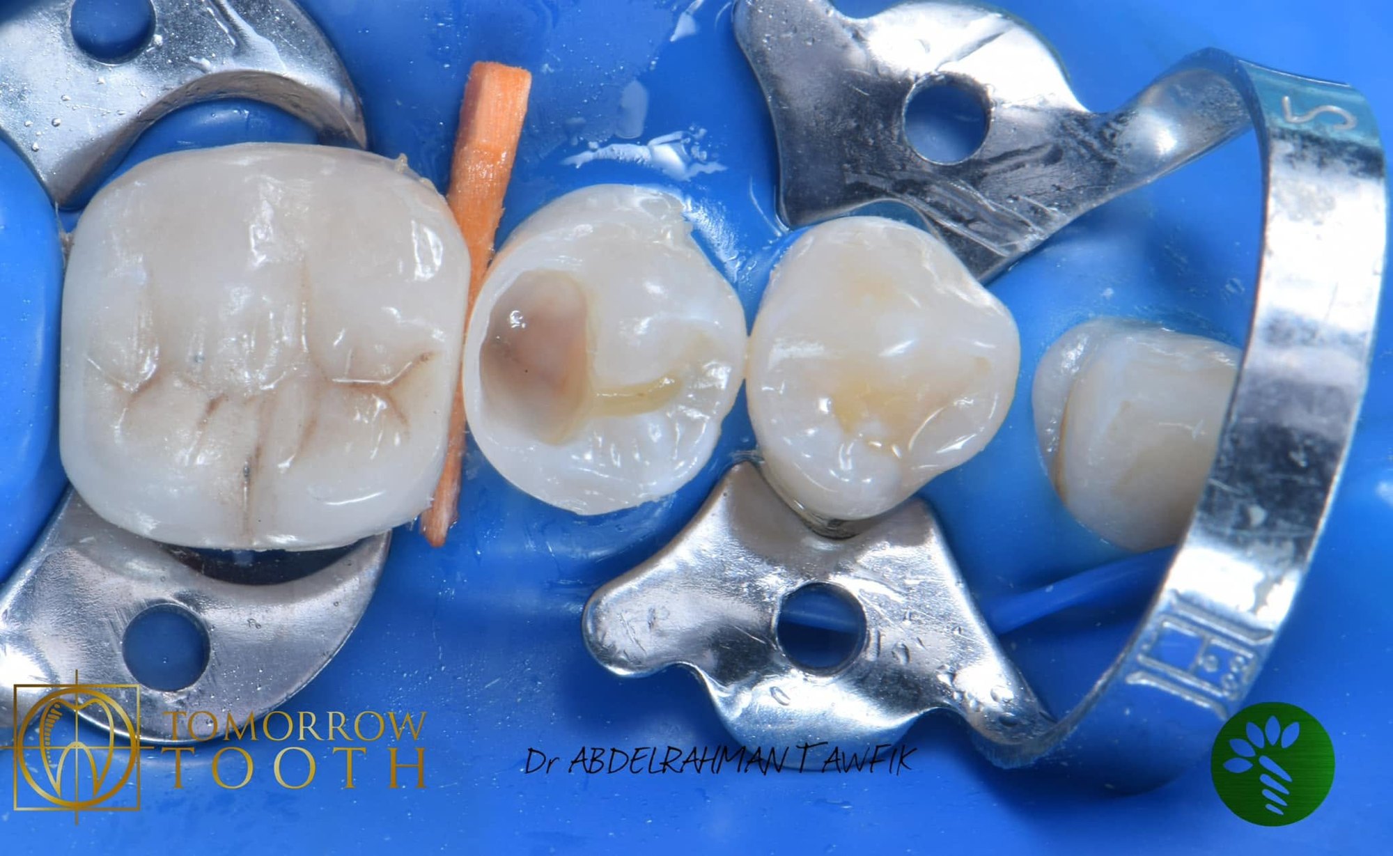
15 Lower molar: proximal wall with TOR VM sectional matrix and Bioclear ring (snowplow) 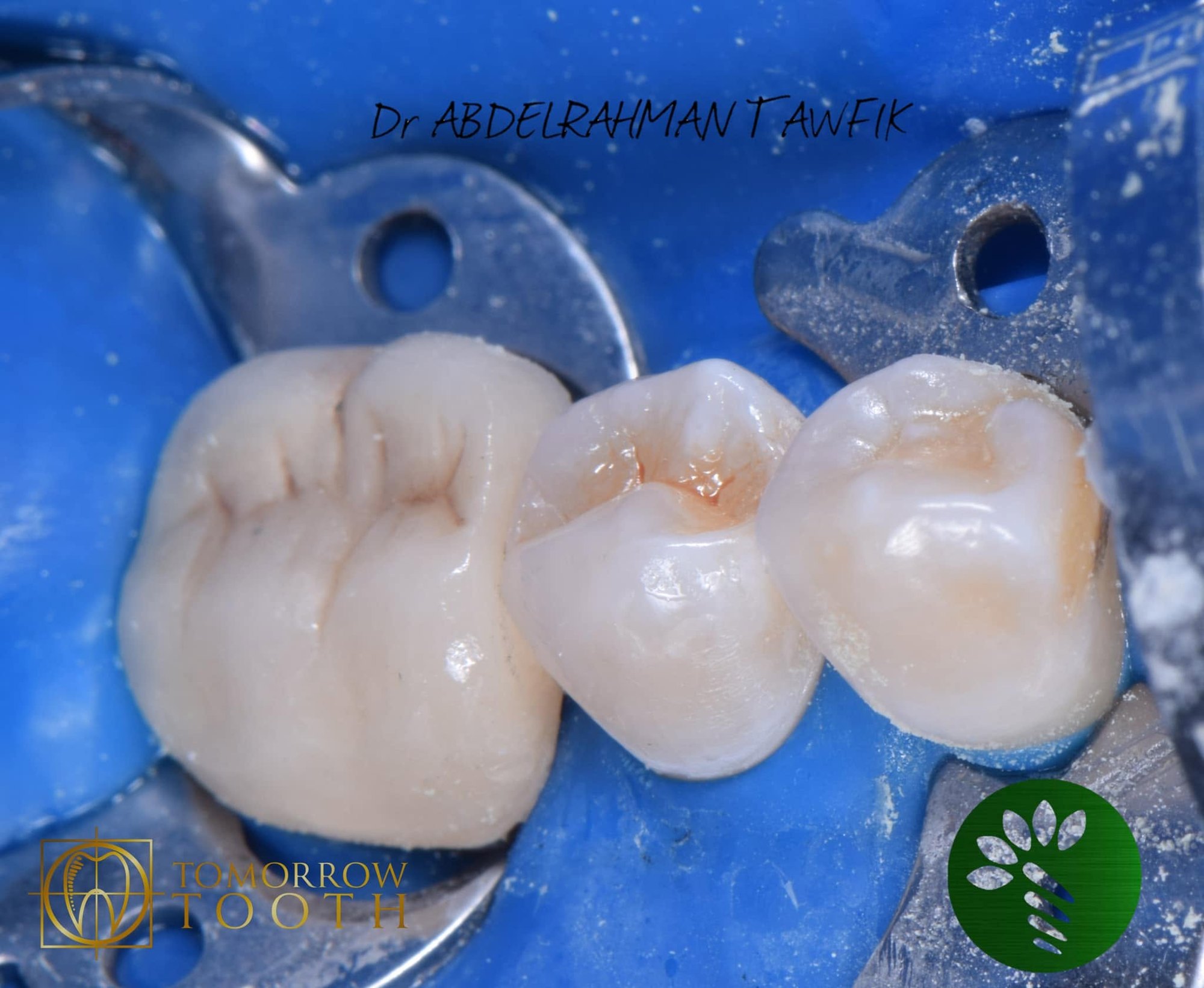
16 Lower molar: occlusal anatomy under rubber dam 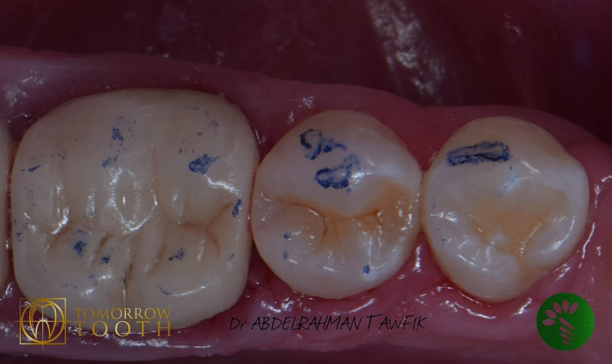
17 Lower molar: occlusion check 18 Lower molar: 7-year follow-up 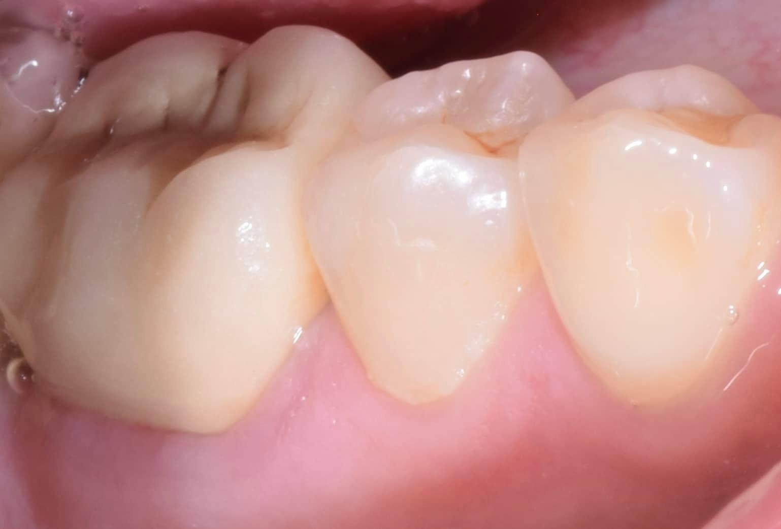
19 Lower molar: 7-year follow-up 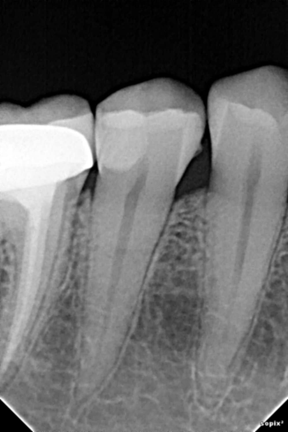
20 Lower molar: 7-year follow-up radiograph
Two teeth, two strategies: 7-year follow-up
- Tooth 1: maxillary left first premolar (#24). Pre-endodontic build-up, root canal treatment and direct cuspal-coverage composite (direct overlay).
- Tooth 2: mandibular molar. Partial (selective) caries removal to preserve pulp vitality, then a direct composite with the proximal wall built first.
Tooth 1: maxillary first premolar
- Deep proximal caries extending subgingivally, with pulp involvement.
- Rubber dam isolation.
- Pre-endodontic build-up with deep margin relocation: Bioclear Biofit matrix, Bioclear Diamond Wedge and Bioclear Twin Ring.
- Snowplow technique at the cervical floor: an uncured layer of flowable composite, with the paste composite pushed into it and both light-cured together.
- Root canal treatment, obturation with a resin-based sealer.
- Definitive direct composite with cuspal coverage (direct overlay).
Tooth 2: mandibular molar
- Deep caries close to the pulp, vital pulp, no signs of irreversible pulpitis.
- Multiple-tooth rubber dam isolation.
- Partial (selective) caries removal: peripheral dentine and enamel margins cleaned to hard tissue, soft dentine left over the pulp to avoid exposure.
- Proximal wall built first with the snowplow technique, using a TOR VM sectional matrix and a Bioclear ring.
- Occlusal anatomy built up, occlusion checked, finished and polished.
Materials
- 3M Filtek Z350 XT composite
- 3M Scotchbond adhesive
- Resin-based endodontic sealer (tooth 1)
Rationale
- Build-up before endo: a predictable rubber-dam seal, a stable reference for working length, and a sealed chamber between visits. Relocating the deep margin follows the deep margin elevation principle, with 95.9% survival reported up to 12 years (Bresser et al., 2019).
- Direct overlay instead of a crown: preserves sound tooth structure. In a randomized trial on endodontically treated premolars, failure rates of direct composite were not significantly different from full crowns (Mannocci et al., 2002). Bruxism is a major risk factor, so case selection matters (Abu-Awwad et al., 2025).
- Coronal seal: the quality of the coronal restoration strongly influences periapical health (Ray & Trope, 1995).
- Partial caries removal: avoiding pulp exposure is the key to keeping the pulp vital. International consensus recommends selective removal in deep lesions (Schwendicke et al., 2016). In a 5-year randomized trial, restorations placed over soft dentine survived as well as those after stepwise excavation (Jardim et al., 2020).
- Longevity: posterior composites show annual failure rates of 1.8% at 5 years and 2.4% at 10 years (Opdam et al., 2014).
Clinical tips
- Rubber dam at every step, including multiple-tooth isolation when restoring adjacent teeth.
- In deep vital teeth, protect the pulp: clean the periphery, not the floor.
- Build the proximal wall first, then the occlusal anatomy.
- Snowplow at the cervical floor for adaptation.
- Use cuspal coverage when the walls are thin.
- Check the occlusion carefully, and recall regularly.
References
- Opdam NJ, et al. Longevity of posterior composite restorations: a systematic review and meta-analysis. J Dent Res. 2014;93(10):943–949.
- Mannocci F, et al. Three-year clinical comparison of survival of endodontically treated teeth restored with either full cast coverage or with direct composite restoration. J Prosthet Dent. 2002.
- Abu-Awwad M, et al. Direct restorations versus full crowns in endodontically treated molar teeth: a three-year randomized clinical trial. J Dent. 2025;156:105699.
- Bresser RA, et al. Up to 12 years clinical evaluation of 197 partial indirect restorations with deep margin elevation in the posterior region. J Dent. 2019;91:103227.
- Ray HA, Trope M. Periapical status of endodontically treated teeth in relation to the technical quality of the root filling and the coronal restoration. Int Endod J. 1995;28(1):12–18.
- Schwendicke F, et al. Managing carious lesions: consensus recommendations on carious tissue removal. Adv Dent Res. 2016;28(2):58–67.
- Jardim JJ, et al. Restorations after selective caries removal: 5-year randomized trial. J Dent. 2020;99:103416.
احجز موعدك
اترك بياناتك، وسيتواصل معك فريقنا لتحديد موعد الزيارة، سواء كنت داخل مصر أو قادمًا من خارجها خصيصًا للعلاج.